
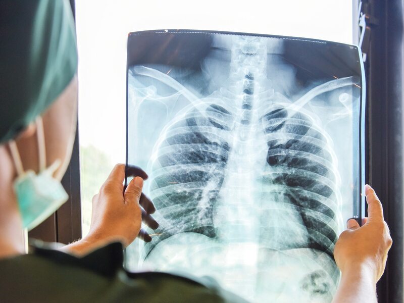
An x-ray of the lungs is a painless examination that is used, for example, at the stage of diagnosing lung diseases. X-rays (X-rays) until recently formed the basis of radiation diagnostics and are still indispensable at the stage of preventive examinations and diagnosis of developing diseases, incl. inside the chest.
- How often should a chest x-ray be done prophylactically?
- What is a chest x-ray?
- How is a chest x-ray performed?
- What diseases can be detected by a chest x-ray?
- What symptoms should prompt a lung x-ray?
- Can anyone do a chest x-ray?
A chest x-ray (chest x-ray) should not be taken too often, due to the adverse effects of high doses of x-rays on the body. A chest x-ray allows you to evaluate the condition of the lungs, heart, and bones of the chest. This examination is carried out not only for diagnostic purposes, but is also an important element of preventive examinations. In addition, chest x-rays are performed to monitor the progress of chronic diseases. This is especially important for tobacco smokers and those at risk of developing occupational diseases associated with working in hazardous conditions.
A chest x-ray can detect various abnormalities in the lungs, heart muscle, and chest wall. This is done, for example. in patients suffering from dry or wet cough for a long time, shortness of breath appears, as well as noises and wheezing in the lungs.
How often should a chest x-ray be done prophylactically?
A chest x-ray is one of the most commonly performed imaging studies. A chest x-ray should be performed prophylactically every two years in non-smokers and once a year in those at increased risk of lung disease.
Indications for more frequent X-ray examinations of the lungs are also, for example. emphysema and chronic obstructive pulmonary disease. In addition, x-rays can diagnose lung cancer, pneumonia, or tuberculosis.
What is a chest x-ray?
X-ray examination of the chest organs is painless and does not cause discomfort. It also does not require special training. X-ray is carried out on modern diagnostic equipment. X-ray imaging is possible due to X-rays that are absorbed by body tissues. Bones absorb X-rays the most, so X-ray examination is one of the main elements in the diagnosis of orthopedic diseases. This is done, for example. in patients with degenerative changes, in patients with injuries of the joints of the arms and legs, as well as in patients with scoliosis and other diseases of the spine.
The soft tissues that make up the body’s organs, such as the lungs and heart, show up as dark spots on an x-ray.. Currently used X-ray equipment allows minimizing the negative impact of X-rays on the body. However, chest x-rays and other x-ray studies are performed only on clear indications from a doctor.
You need to get a referral from a doctor to have an x-ray.
How is a chest x-ray performed?
A chest x-ray takes a short time. Before the examination, the patient must remove clothing from the examined part of the body and jewelry. Then he is instructed how to position himself in front of the x-ray machine. X-rays of the lungs can also be taken with the patient lying down.
Before starting the X-ray machine, the patient must take a deep breath. The lungs may be deflated after a chest x-ray. During the examination, it is necessary to protect the thyroid gland and other sensitive areas of the body, such as around the testicles, with special screens through which X-rays do not penetrate.
Chest x-ray can be done in different positionsThat is:
-
in the posterior-anterior projection (ZP),
-
in the anterior-posterior (AP) projection,
-
in lateral projection
-
in an oblique view.
Most often, chest x-rays are performed in the posterior-anterior and anterior-posterior projections.
What diseases can be detected by a chest x-ray?
With a chest x-ray, your doctor can evaluate the condition of your lungs and other chest organs. If a chest x-ray of a patient shows that, for example, lung cancer is developing, additional studies should be carried out, i.e. computed tomography studies.
Other conditions that can be diagnosed on a chest x-ray include: pneumonia, lung tumors, tuberculosis, emphysema, as well as heart failure and other cardiac diseases, mediastinal lymph node enlargement, and retrosternal goiter.
What symptoms should prompt a lung x-ray?
An x-ray of the lungs, if it is not part of a health prophylaxis, is performed when the patient has symptoms that indicate pulmonary or cardiac diseases. A chest x-ray is usually done when the patient:
-
symptoms suggestive of lung cancer, including chronic cough, chest pain, hemoptysis, weight loss,
-
shortness of breath appeared
-
exacerbation of symptoms of chronic lung disease,
-
symptoms appear that indicate a violation of the heart, for example, pain behind the sternum, burning in the chest, weakness, arrhythmias,
-
there are recurrent pneumonias,
-
there are symptoms indicating changes in the pleural cavity,
-
there are defects in the development of breast tissue.
An indication for lung radiography is also a suspicion of the presence of cancer metastases in the lungs.
If you experience the following symptoms: chest pain and tightness, shortness of breath, shortness of breath with little exertion, fatigue, coughing up bloody sputum, you should contact your doctor as soon as possible, who will decide whether an X-ray is needed.
Before the patient hears the diagnosis, the radiologist must describe the chest X-ray, which looks for abnormalities not only in the lungs, but also in the lungs, among other things. diaphragm, ribs, heart and lymph nodes.
Can anyone do a chest x-ray?
There are contraindications to X-ray examination. If it can be avoided - the absence of an examination will not pose a threat to health and life - it is not carried out for pregnant women. X-ray studies are also not carried out without clear indications in newborns and young children.
Before performing chest x-rays in patients who are particularly susceptible to adverse x-ray exposure, it is necessary to consider not only the benefits, but also the risks associated with x-rays.
Sources:
- Schaefer-Prokop K., Dixon A., Radiological Diagnostics, Urban&Partner Publishing House, Wroclaw 2017
- Ptak, J. et al., Imaging studies in pneumonia in adults, Pneumonology and Allergology, Poland, 2011, Volume 79, Number 1, Pages 57–66.
Source: Wprost

I have worked in the news industry for over 8 years. I have a deep understanding of how the news industry works and how to get information out to the public. I am also an author at Daily News Hack, where I mostly cover health news. I have a keen interest in health and fitness, and I firmly believe that knowledge is power when it comes to taking care of your body. I want to help people live healthier lives by sharing my knowledge with them, and LinkedIn is the perfect platform for me to do that.